After two years convening remotely through computer screens, thousands of eager physicians, researchers, and other professionals in the cancer space converged on New Orleans—like T cells storming a tumor—for the 2022 annual meeting of the American Association for Cancer Research (AACR22).
As one speaker captured the mood, “It’s so good to be here in vivo, not just in silico.”
Immunotherapy is now an established pillar of cancer care, thanks to checkpoint inhibitor and adoptive cell therapies that tap into the power of T cells, but the limits of currently approved T approaches are becoming clear. Alone, they still don’t work for most people or types of tumors. By diving deeper into the basic biology and identities of T cells though, we might develop better ways to utilize them against all cancers.
“We really don't understand fully what kinds of T cells or what kind of immune cells are playing a role in cancer therapy, and what kind of dysfunction might underlie the limited responses we've seen in many patients,“ according to E. John Wherry, Ph.D., an associate director of the CRI Scientific Advisory Council who was first funded by CRI as a postdoctoral fellow in the early 2000s.

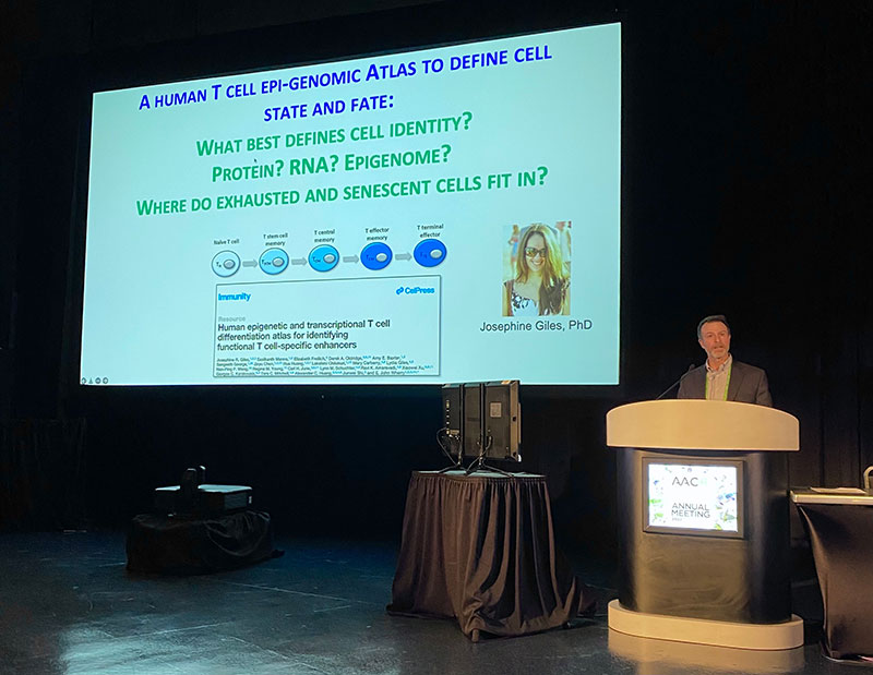
E. John Wherry, Ph.D., at AACR22. Photo by Arthur Brodsky
Josephine Giles, Ph.D., a CRI-Mark Foundation Fellow in Wherry’s lab at the University of Pennsylvania School of Medicine, led the creation of a T cell atlas that defines various T cell states and identities based on what genes they’re expressing and the accessibility of their genomes (epigenomics). That baseline information then provides better context for the types of T cells seen in the tumors of those with cancer, and could reveal how to nudge them in the right direction therapeutically.
With this atlas, they classified how exhaustion differs from senescence in T cells, and found that exhausted T cells predominate in most cases of melanoma. Building on that, Wherry’s team developed tools to monitor how T cells respond to DNA damage, demonstrating the importance of DNA repair and maintaining genome integrity when it comes to persistent T-cell responses and successful checkpoint immunotherapy.
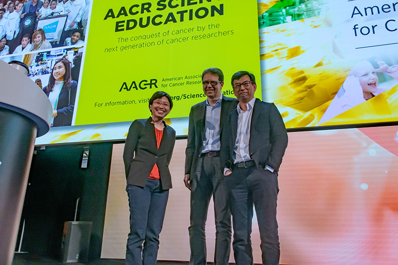
Turning to cell therapies, Cassian Yee, M.D., of the University of Texas MD Anderson Cancer Center, chaired a session with Michel Sadelain, M.D., Ph.D., of Memorial Sloan Kettering Cancer Center, and Yvonne Chen, Ph.D., of the University of California, Los Angeles (UCLA) Jonsson Comprehensive Cancer Center.

Drs. Yvonne Chen, Michel Sadelain, and Cassian Yee at AACR22. Photo by Arthur Brodsky
Together, Chen, Sadelain, and Yee explored sophisticated strategies for expanding the benefits of these incredibly promising cell therapies to more patients, and how we might go about developing successful cell therapy platforms for all types of cancer.
Chimeric antigen receptor (CAR) T cells have benefited many with advanced blood cancers like leukemia and lymphoma by targeting CD19 and myeloma by targeting BCMA. However, there are many problems that still need to be overcome to improve their value for patients. Manufacturing hurdles aside, toxicity issues remain, and relapse can sometimes occur even after an impressive response initially. One cause of relapse is antigen escape, where cancer cells lose or diminish their levels of the target protein to avoid detection by T cells.
To address this, Chen, an inaugural CRI Lloyd J. Old STAR (Scientist TAking Risks), designed CAR T cells targeting both the CD19 and CD20 antigens. Of the eight people with either leukemia or lymphoma who received these CAR T cells in the Phase 1 trial, seven had complete responses, and the longest response remains ongoing at two years and counting. One person who saw their disease disappear had four prior lines of treatment, including CD19 and CD20 therapies, and their cancer cells expressed low levels of CD19, the dominant target. As for the person who didn’t respond, they appeared to have small subpopulations of cancers cells that had already lost both CD19 and CD20, thus making them invisible to the CAR T cells.
_1415.jpg)
Dr. Yvonne Chen at AACR22. Photo by Arthur Brodsky
These CAR T cells were also safe, likely due to the lower dose compared to currently approved CAR T cell therapies: 50 million versus 1 billion cells. Only one person needed a remedy for cytokine release syndrome, a potential side effect of CAR T therapy. This patient had elevated concentrations of inflammation-related cytokines prior to the therapy, and they remain relapse-free.
“This is really encouraging,” said Chen, “because it shows us that with CAR T cells, we can achieve very high efficacy without triggering severe CAR T cell-related toxicity.”
CAR T cell persistence, or lack thereof, is another cause of relapse. If CAR T cells don’t last long enough within the patient, the cancer can return. Here, a seeming paradox exists: if T cells bind a target too strongly, they can become exhausted (in some cases). By adding amino acids to “twist” the structure of CARs, Chen identified certain conformations that protected against T cell exhaustion, and improved cytokine production and performance, with both CD19-targeting CARs in blood cancer and GD2-targeting CARs in neuroblastoma. Mixing binding elements from strong and weak CARs conferred synergistic benefits in some combinations, enhancing both the speed and robustness of CAR T cell responses.
Sadelain, a cell therapy pioneer who designed the first CAR T cells used in human trials and serves on the leadership of the CRI Anna-Maria Kellen Clinical Accelerator, highlighted his latest advances, including synthetic immune receptors beyond CARs. One approach used an “if/better” system supplied with an additional receptor—a chimeric co-stimulatory receptor (CCRs)—that supports CAR T cell persistence and improves their antigen sensitivity. Sadelain also shared a recent peek into his HLA-independent T (HIT) cell receptor technology, which incorporates a CAR—and its specialized binding capabilities—into the natural T cell receptor complex, in order to mimic the natural signaling and processing mechanisms as much as possible. These HIT receptors offered the most sensitivity and triggered “more complete” T cell activation. In mice, they could eliminate cancer cells with as few as 200 CD19 molecules on their surface.

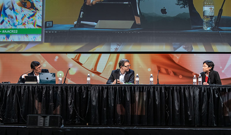
Drs. Cassian Yee, Michel Sadelain, and Yvonne Chen at AACR22. Photo by Arthur Brodsky
In contrast to blood cancers where the same targets—CD19, CD20, BCMA—are found on almost all of the cancerous B cells in patients, solid tumors have no such universal target to go after. That’s why there aren’t any approved cell therapies for solid tumors yet. Some types, such as advanced melanoma, often have many mutations that provide valuable targets for T cells.
With CRI support in the early 2000s, Yee and his colleagues took advantage of naturally-occurring T cells that recognize and infiltrate melanomas, and provided some of the initial proof that adoptive cell therapy could work for solid cancers that collectively are responsible for the vast majority of cancer’s toll. At AACR22, Yee, now a CRI-Chordoma Foundation CLIP Investigator, outlined the culmination of his experience, in the form of a framework that could guide the creation of effective T cell therapies for all cancers.
It starts with supplying patients with the optimal T cells, in this case by generating long-lasting memory T cells. Then, of course, we need to be able to identify the best targets for T cells. Yee’s new antigen discovery platform yielded more than one hundred high-value, T cell-stimulating targets across more than 30 tumor types covering more than 80% of cases. As this immunopeptidome atlas is further populated and refined, it could aid the discovery of targets in those with rare tumors like chordoma.
In addition to the harder task of identifying targets in solid cancers, there is another, even more formidable task that’s unique to these tumors: the need to overcome barriers within the tumor microenvironment for either cell therapy or checkpoint immunotherapy to succeed. And to truly appreciate the tumor microenvironment’s influence in cancer immunotherapy we must bring to center stage, to their rightful place alongside T cells, another group of immune cells: myeloid cells.
Myeloid cells come in many diverse forms and subsets, and when it comes to adaptive immunity they are similar to generals in an army. They tell the T cell soldiers what and when to attack, and when to stand back. T cells will always remain a very necessary part of anti-cancer immunity, but as Genentech’s Ira Mellman, Ph.D., put it, “They would be nowhere were it not for the myeloid cells.”
In one of the most exciting sessions of the conference, Mellman—the recipient of the 2022 AACR-CRI Lloyd J. Old Award in Cancer Immunology—explored the roles of these important immune cells, along with Miriam Merad, M.D., Ph.D., and Brian D. Brown, Ph.D., of the Icahn School of Medicine at Mt. Sinai, as well as Arja Ray, Ph.D., of the University of California, San Francisco.
Their collective message: a better understanding of how myeloid cells mediate immunity within tumors will pave the way for the next transformative breakthroughs in cancer immunotherapy.
_1245.jpg)
Dr. Ira Mellman at AACR22. Photo by Arthur Brodsky
Myeloid cells, in Mellman’s estimation, “make the decision as to whether a tumor is going to adopt an immune profile that will be susceptible to checkpoint inhibition.” In clinical trials, his group identified PD-L1 expression by myeloid cells as an important factor linked to patient responses.
Dendritic cells are especially important. They are the primary antigen-presenting cells and express co-stimulatory receptors that enhance T cell activation, but they also express the majority of PD-L1 within tumors, which can prematurely shut down T cells in certain conditions. Knocking out PD-L1 expression just in dendritic cells was as effective as systemic inhibition, both of which were more effective than just blocking macrophage PD-L1 expression.
Data from the lab as well as a trial using dual checkpoint blockade against the PD-1/PD-L1 and TIGIT pathways in lung cancer suggested that interactions between myeloid cells and the “back ends” of the TIGIT-targeting antibodies were crucial for its effectiveness. Lastly, Mellman showed how, even when genetically identical tumor cells were injected into the same mouse, the “soil” of the tumor microenvironment and the myeloid cells there determined the immunological fate of those tumors and their responsiveness to immunotherapy.
_1297.jpg)
Dr. Miriam Merad at AACR22. Photo by Arthur Brodsky
Miriam Merad, M.D., Ph.D.—a CRI Scientific Advisory Council member who currently sponsors CRI fellow Nelson Lamarche, Ph.D., in her lab—discussed work led in collaboration with CRI CLIP Investigator Thomas Marron, M.D., Ph.D., as part of TARGET, The neoAdjuvant Research Group to Evaluate Therapeutics. In their trial, people with early-stage liver cancer were treated with checkpoint immunotherapy prior to surgery, and this neoadjuvant approach resulted in elimination of at least half the tumor by the time of resection in 28% of patients.
Through deep analysis of the samples, they revealed the importance of two different T cell subsets, effectors for killing and follicular helpers for support. Additionally, they defined a state that’s adopted by mature dendritic cells after they uptake tumor antigen and become activated. These specialized immune cells actively engage T cells, forming tight clusters that appear to promote T cell priming, activation, and survival. Merad emphasized the need to improve our understanding of the niches these cells create and manage, and how exactly they promote effective immunotherapy. Being able to follow cells and other factors over time would be even more helpful.
_1221.jpg)
Dr. Brian Brown at AACR22. Photo by Arthur Brodsky
To that end, Brown, a CRI Technology Impact Award recipient who works closely with Merad at Mt. Sinai, discussed cutting-edge tools created with CRI funding that enable him to apply CRISPR genetic engineering screens in a novel way, in order to analyze not only genes within cells, but also the factors outside cells in the tumor microenvironment. Applying this multiplex screen to lung tumors, Brown’s project identified two important pathways in cancer cells whose disruption enhanced tumor progression, but in distinct and telling ways, especially with respect to how each type remodeled the architecture of the tumor microenvironment.
For the first pathway, Brown showed that loss of Socs1 activity promoted “hot” tumors that were susceptible to immunotherapy-induced immune responses, whereas the loss of the ability to respond to TGF-b, the second pathway, promoted “cold” tumors that were able to keep T cells at bay. This signature was observed in several other types of cancer, such as breast, ovarian, and pancreatic that are often immunologically cold and unresponsive to current checkpoint immunotherapies.
To close this session, CRI Fellow Arja Ray, Ph.D., showcased another new system to define the states of immune cells within tumors. T cell activation is a complex process, and Ray and his colleagues wanted to be able to measure a T cell’s potential for re-activation over time in as precise and directed a fashion as possible. In developing this approach, they were able to accurately track the cell’s progression from “poised” for activation, to acute activation, and finally to chronic activation, exhaustion, and quiescence. Notably, they identified CD81 as a reliable marker of the T cells that were best at mediating tumor regression, and whose presence predicted survival across different types of cancer.
_1348.jpg)
CRI Fellow Arja Ray, Ph.D. Photo by Arthur Brodsky
The tumor microenvironment and myeloid cells popped up in other prominent sessions, too, including a plenary featuring the University of Chicago’s Thomas F. Gajewski, M.D., Ph.D., and Genentech’s Shannon Turley, Ph.D.
Gajewski, the recipient of the CRI’s 2017 William B. Coley Award, spoke about three sources of patient heterogeneity that influence the immunological nature of their tumor microenvironments: (1) DNA repair in tumor cells, (2) the composition of our bacterial microbiomes, and (3) natural genetic differences linked to familial lupus that also impact cancer immunity. With a new system that enables his team to isolate the impact of defined gut microbiota that can be passed down generationally, they uncovered bacteria-related metabolites in the blood that were linked to immunotherapy responsiveness. Regarding automimmune diseases, Gajewski called that field, “a new land through which we (cancer immunologists) should search for therapeutic targets,” noting that CTLA-4 and PD-1 were both mentioned in autoimmunity research prior to cancer. As he demonstrated during his talk, each of these mechanisms involves the regulation of myeloid cells at some level.
Turley, a former CRI fellow who received the Frederick W. Alt Award at a 2017 CRI awards ceremony, focused on cells called fibroblasts found within tumors. Though not immune cells, they are an important part of all tissues, including the tumor microenvironment, where they synthesize and maintain the extracellular matrix that acts as scaffolding. Specifically, she found that, in contrast to immune response-activating dendritic cells, activated fibroblasts known as myofibroblasts cluster with T cells in a way that negatively impacts natural immunity and immunotherapy responses in pancreatic cancer. Turley also discussed her team’s creation of a tool to analyze the diversity of fibroblast populations and lineages across all tissues, under both healthy and disease conditions. Thus far, she’s focused on characterizing the myofibroblast subsets that have been implicated in a variety of other inflammatory diseases beyond cancer, including arthritis, colitis, pulmonary fibrosis, and COVID-19.
_1320.jpg)
Dr. Susan Kaech at AACR22. Photo by Arthur Brodsky
Susan M. Kaech, Ph.D., of the Salk Institute, also chaired a session where she and Lydia Lynch, Ph.D., of Harvard Medical School, both CRI CLIP Investigators, presented work in the emerging area of cancer immunometabolism. In both these areas, great consideration was given to the nature of the tumor microenvironment as a whole.
Kaech’s work provided yet another connection to myeloid cells via metabolism. During tumor development in the lungs, macrophages get co-opted and metabolically reprogrammed by cancer cells to become more anti-inflammatory. In this state, the macrophages became much more dependent on lipids in the tumor microenvironment. Consequently, as the macrophages consumed more lipids, lipid metabolism decreased in T cells, negatively impacting their activity. Fortunately, Kaech found that blocking the PPAR gamma pathway could cause macrophages to store rather than consume the lipids, and this resulted in delayed tumor progression.
_1307.jpg)
Dr. Lydia Lynch at AACR22. Photo by Arthur Brodsky
Lynch’s work defined how obesity, which is linked to increased risk of cancer as well as infection, impacts T cells as well as natural killer (NK) immune cells, in part through the elevated levels of lipids that Kaech recognized. Lynch found that while both calorie reduction and a ketogenic diet reduced circulating levels of glucose and insulin, only the former reduced lipid levels and tumor growth. Additionally, her studies linked obesity to NK cells that were “metabolically paralyzed” and associated with an overdependence on lipid metabolism. In contrast, however, Lynch also observed in human endometrial cancer that immune-suppressing cells like regulatory T cells and gamma delta T cells thrive in the lipid-rich tumor microenvironment, enabling them to support tumor growth and survival. As for the source of lipids, compared to palm oil, the animal-derived fats (from butter) were found to be more damaging to T cell metabolism, resulting in more impairment and mitochondrial dysfunction.
Those are the top cancer immunotherapy developments to come out of AACR22, which proved to be a very exciting and long-awaited conference. With experts from all disciplines spreading the latest advances in cancer immunology among each other, there was a palpable and rejuvenating sense of progress on display, one that will hopefully accelerate discovery from the next wave of laboratory experiments and clinical trials, ultimately leading to more effective cancer treatments and even cures for more people.